Fmge examinations in January 2024 were not good for most students.
The results were disappointing for most of the students. 75 percent students did not fare well and failed to qualify.
What were the reasons? Hadn’t the students prepared well. Definitely there must have been reasons for failing to qualify. Routine methods of preparation failed.
The FMGE Examination has undergone a lot of change. Newly conducted FMGE Examinations were a bit different and more clinically oriented.
Many FMGE students had not taken their clinical postings seriously. There are some things to learn in OPD and Ward rounds especially Image based questions.
There is a need for high yield content and best possible source of information about latest developments.
The students need to focus on all subjects, all important topics, Best Possible Explanations, High yield Points, Extra Edge and Image Based Text wherever applicable.
Where from do the Majority of Students Prepare:
- FMGE Students Maldives
- FMGE Student India
- FMGE Student China
- FMGE Student Russia
- FMGE Student Bangladesh
- FMGE Student Philippines
- FMGE Students Ukraine
The Factors for not doing well are:
- Not Good sources of information.
- Relying on Advertisements and packages without consultation properly
- Studying from unnecessary video channels
- Not focusing on Standard Text materials
- Looking for short cuts to success
Reading from standard text books must be encouraged. Main topics from books like Harrisons medicine, Bailey surgery, Robbins Pathology, should be encouraged.
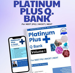
The Platinum Q Bank is a new addition to the Preparation for FMGE Students. New Students preparing for FMGE and NEET PG can benefit from the book as the book provides a Good q bank and the yield for exams can be high. The Questions are clinically oriented and some latest topics and image based questions have been added. The Book needs two to three revisions.
Examples of Questions from Platinum Q Bank (With Permission of Author)
Q: A Child suffered an injury after birth which was caused by forceful downward traction of shoulder with lateral displacement of head to the other side causing the Hanging of arm by the side and Extension of elbow. Most likely deformity is:
- Erb’s Palsy
- Klumpkes Palsy
- Pulled Elbow
- Saturday night palsy
Ans A Erb’s Palsy
Erb’s point: union of six nerves namely C5, C6, Suprascapular nerve, Nerve to subclavius, Anterior and posterior division. Damage to it results in Erb’s paralysis. (Policeman’s tip) Erbs Palsy: Lesion of upper trunk of Brachial plexus(C5, C6) caused by forceful downward traction of shoulder with lateral displacement of head to the other side.
The deformity causes:
- Hanging of arm by the side.
- Extension of elbow
- Pronation of forearm
- Flexion of wrist.
Q: A child has a biochemical deficiency of galactose-1-phosphate uridyltransferase. Likely features would be:
- Alkalosis , glycosuria, aminoaciduria and hypoglycemia
- Acidosis , glycosuria, aminoaciduria and hyperglycemia
- Alkalosis , glycosuria, aminoaciduria and hyperglycemia
- Acidosis , glycosuria, aminoaciduria and hypoglycemia
Ans D Acidosis , glycosuria, aminoaciduria and hypoglycemia
Galactosemia:
Galactosemia is an autosomal recessive disease caused by deficiency of galactose-1-phosphate uridyltransferase. Clinical manifestations are most striking in a neonate who, when fed milk, generally exhibits evidence of liver failure (Hyperbilirubinemia, disorders of coagulation, hypoglycemia), disordered renal tubular function (acidosis, glycosuria, aminoaciduria),
Q: A 44 year has peritonitis. Gram positive cocci in chain with catalase negativity were isolated. Organisms causing the same are gamma-hemolytic that grow on bile-esculin agar. Most likely organisms are:
- Streptococci
- Staphylococci
- Meningococci
- Enterococci
Ans D Enterococci
Enterococcus
- Causes Urinary tract and biliary tract infections Endocarditis rarely but life threatening.
- Characteristics of Enterococcus faecalis are gram-positive cocci in chains. Catalase-negative.
- Laboratory Diagnosis of Enterococcus faecalis is gram-stained smear and culture. Alpha-, beta-, or nonhemolytic colonies in blood agar. Grows in 6.5%.NaCL and hydrolyzes esculin in the presence of 40% bile. Serologic tests not useful.
Q: A 33 year old boy presented with a swelling in his right leg. X ray demonstrated a mass arising in the distal right femur. X-ray from the lesion showed features of Codman’s triangle. The diagnosis is:
A. Osteosarcoma
B. Chondrosarcoma
C. Aneurysmal bone Cyst
D. Osteoclastoma
Ans A Osteosarcoma
Osteogenic sarcoma is a highly malignant primary bone tumor. Here tumor cells invariably form a neoplastic osteoid, bone, or both. It arises from a common multifactorial mesenchymal tissue and hence the tumor could be either fibroblastic, osteoblastic or chondroblastic. Sunray Spicules and Codman’s triangle are an important distinguishing feature
Q: A 13 year old patient is labeled as having Bergers Disease. It is characteristically a Type of:
- Nephropathy
- Neuropathy
- Lymphadenopathy
- Retinopathy
Ans A Nephropathy
IgA Nephropathy (Berger disease)
- IgA nephropathy is more common in males than in females (2:1). Patients either present with an episode of gross hematuria or are found to have microscopic hematuria on routine examination.
- While the gross hematuria lasts, renal function usually remains relatively normal and proteinuria minimal (<1 g/24 hr).
- Normal serum levels of C3 in IgA nephropathy help to distinguish this disorder from post streptococcal glomerulonephritis.
- Microscopic Examination reveals the mesangial widening and proliferation which on immunofluorescence reveals the presence of the mesangial deposition of IgA usually with C3 and properdin. Mesangioproliferative glomerulonephritis is seen more commonly than focal proliferative and crescentic (least) glomerulonephritis.
Q: A 12 year old child had Fever with Abdominal pain. Meckel's diverticulum is the most likely Possibility. It is best diagnosed by:
A. Thallium Scanning
B. Ultrasound
C. CT scan
D. Tc99 pertechnetium scan
Ans D Tc99 pertechnetium scan
Diagnosis of a Meckel's diverticulum possessing gastric mucosa can be made using 99mTc-pertechnetate radioisotope scanning. This isotopic compound is readily taken up by gastric mucosa. Pharmacologic intervention with pentagastrin or a histamine H 2 receptor blocker can significantly enhance the detection of a Meckel's diverticulum by enhancing uptake and inhibiting intraluminal release, respectively, minimizing false negative results. 6 Angiography, ultrasonography, and intestinal contrast studies have also been reported as means of preoperatively diagnosing a Meckel's diverticulum, but their use is not routine in these patients. The Tc 99 scan is yet to be surpassed by any other modern investigative procedure.
Image Based Questions
Q: A young girl has a lesion as shown below. The most likely diagnosis of below shown condition is:

- Neurofibroma
- Ephelid
- TENS
- Port Wine Stain
Ans D Port wine Stain
The Most likely lesion in figure is:
Port-wine stain (nevus flammeus): Unlike a strawberry nevus this is present at birth and may be very disfiguring, as it becomes darker and increasingly nodular with age. It persists. Persists (P for P)
Q: CT Scan demonstrates cholelithiasis. Most common types of stones in cholelithiasis are:

- Magnesium stones
- Struvite stones
- Cysteine stones
- Cholesterol stones
Ans D Cholesterol stones
Gallstones are the most common biliary pathology. Gallstones are classified according to their chemical composition into cholesterol stones, mixed stones and pigment stones. Cholesterol stones consist almost entirely of cholesterol and are often solitary. Mixed stones account for most of gallstones. Cholesterol is the major component. Pigment stones are composed almost entirely of calcium bilirubinate. They are mostly small and multiple. Bile containing cholesterol stones has an excess of cholesterol relative to bile salts and phospholipids, thus allowing cholesterol crystals to form. Such bile is termed ‘supersaturated’ or ‘lithogenic’.
These Examples of Questions from Platinum Q give you a rough idea for how to get an idea about the Question types asked in Examinations.
Links for Purchasing Platinum Q Bank are :
https://notionpress.com/read/platinum-plus-q-bank-volume-i
https://notionpress.com/read/platinum-plus-q-bank-volume-ii
We hope that this helps you in getting an idea. For Online Q Bank you can directly subscribe to:
https://www.medexamsprep.com/category/package/fmge